Group 3
Amel Slimani (group leader)
- Frédéric CUISINIER (PU-PH)
- Jean Valcarcel (PU-PH)
- Michel Fages (PU-PH)
- Ivan Panayotov (MCU-PH)
- Jean Cédric Durand (PU-PH)
- Alban Desoutter (technical assistant LBN)
- Elias Estephan, Université la sagesse, Liban (PU)
Our scientific objective is to identify new photonic biomarkers for oral diseases, mainly dental caries, periodontal diseases and oral cancer. These pathologies will be studied by different spectroscopic methods and devices. We organize this project according to the methods even if the methods can be combined to increase the specificity and the sensitivity of biomarkers.
-
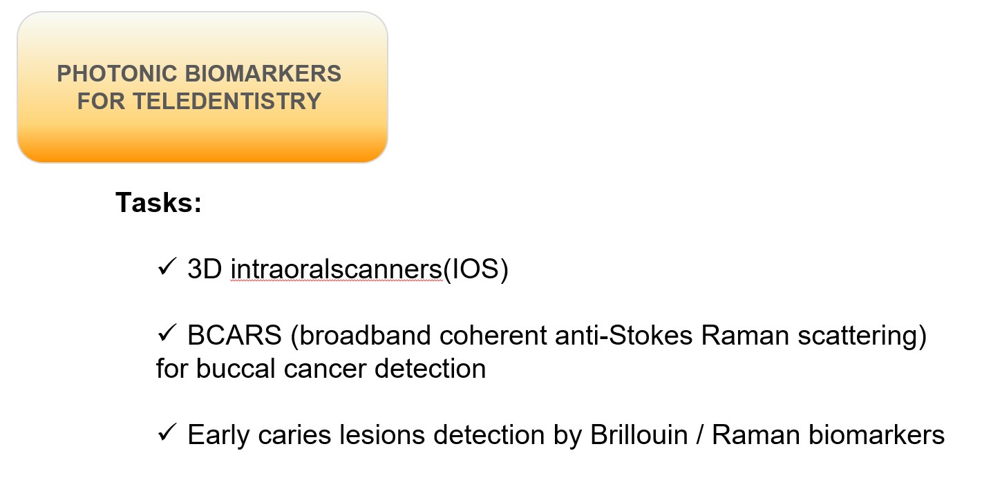
3D intraoralscanners(IOS):
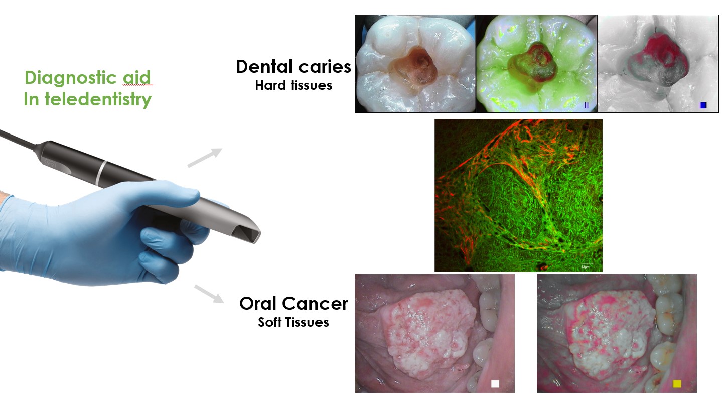
Oral cancer is a significant health problem often identified at later stage. Oral cancers are characterized by a change of color of the tissues as well as oral mucosa thickening. Concerning thickening of tissue, obtaining 3D mesh of oral cavity is an interesting tool to create standardized biomarkers of cancerous lesions. Autofluorescence, color and 3D meshes could be combined to detect oral cancer. We will focus our work to explore the use of the two IOS in order to optimized cancer detection. For this 3D meshes from malignant lesions and sound tissue will be collected. Segmentation and color spectral analyze will lead to identify biomarkers through multivariate statistics. This can be extended to periodontal disease.
-
BCARS to detect the buccal cancer
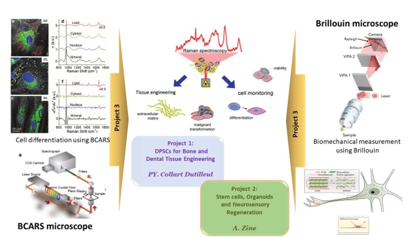
The diagnosis of malignant and cancerous lesions in the oral cavity is the main interest in the development of the new imaging techniques. The malignant tissues make biochemical changes called tumor markers inducing variation in the vibrational spectra. Raman spectra from diseased and healthy tissue are compared for diagnosis. Raman biomarkers are complementary to biopsy and give real-time diagnosis. The first part of the task is to compare the existing standard biopsy analysis with our label free B-CARS signal. The relevant data analysis could detect the modification in tissue with the potential to be used in operating room.
-
Early caries lesions detection by Brillouin / Raman biomarkers
The initial caries lesions are challenging to detect for a dentist and even more in teledentistry. In our laboratory, we have developed a WSLin vitromodel and we used confocal Raman spectroscopy and multiphoton microscopy images to evaluate structural changes in the white spot lesion. The goal of this task is to adapt a new non-invasive biomedical imaging technology based on Brillouin light scattering to measure the biomechanical properties of hard tissues and biomaterials, is the main aim of this project. The molecular analysis of dental tissues using Raman spectroscopy is been used to characterize the mineral phase in synthetic compounds and natural tissues. Our project is to use confocal Raman microscopy coupled with a home-build Brillouin spectrometer. The first step will be to demonstrate the possibility of biomechanical/biochemical simultaneous imaging to obtain. photonic biomarkers for early caries and WSL diagnosis studying in-vitro incipient caries lesions, animal induced periodontal. Comparison with healthy samples will allow statistical analysis to find a biomarker to detect the caries in early level is the final step.
Partnerships:
1- L2C-UMR5221 CNRS: we will continue our strong collaborations through our involvement in the financing and building the BCARS (team Nanobiophonic) and the building of the Brillouin Spectrometer (B. Rufflé, D. Felbacq, team physics of glassy state). We will develop a partnership for colorimetry in teledentistry (F. Geniet, team complex system and nonlinear phenomena).
2- LIRMM: all works of metrology for IOS are discussed and conducted with LIRMM (G. Subsol, ICAR team).
3- LNE: concerning the metrology of IOS, characterization of object and samples tested, we have a collaboration with a team of LNE (Laboratoire National de Métrologie et d’Essai, Nimes).
The press talks about us
About enamel Drapes:
About Brillouin microscopy:
- Midi Libre : https://www.midilibre.fr/2024/07/11/a-luniversite-de-montpellier-un-nouveau-microscope-unique-en-france-12056305.php#:~:text=%C3%80%20l’Universit%C3%A9%20de%20Montpellier%2C%20un%20nouveau%20microscope%20unique%20en%20France,-Abonn%C3%A9s&text=Inaugur%C3%A9e%20le%2027%20juin%20dernier,des%20animaux%20et%20des%20plantes.
- Ecomnews : https://ecomnews.fr/news/montpellier-luniversite-de-montpellier-se-dote-dun-microscope-unique-en-france/
- Tout Montpellier : https://toutmontpellier.fr/news/le-laboratoire-bioingenierie-et-nanosciences-se-dote-dun-microscope-unique-en-france
- La newsletter l’Essentiel Montpellier : https://www.linkedin.com/posts/lessentielmontpellier_microscope-sant%C3%A9-biologie-activity-7212006249247244288-T9e6/?originalSubdomain=fr
- La Marseillaise : https://www.lamarseillaise.fr/societe/le-premier-microscope-brillouin-monte-en-france-BG16458292
- https://www.umontpellier.fr/articles/le-laboratoire-bioingenierie-et-nanosciences-et-le-laboratoire-charles-colomb-inaugurent-le-fonctionnement-dun-microscope-unique-en-france
- https://www.linkedin.com/posts/michael-delafosse_montpellier-activity-7218907077526597633–aLG?utm_source=share&utm_medium=member_ios
Other: